Chest And Abdominal Muscles Diagram ~ Anterior View of the Muscles of the Trunk | ClipArt ETC. See more ideas about muscle diagram, medical anatomy, human anatomy and physiology. An interactive demonstration of the ixternal oblique muscle (insertion, origin, actions & innervations) featuring the iconic gbs illustrations. Muscle performance in neck pain assessment and rehab of the deep. Common chest and abdominal injuries. Learn about each muscle, their locations & functional anatomy.
The abdomen (colloquially called the belly, tummy, midriff or stomach) is the part of the body between the thorax (chest) and pelvis, in humans and in other vertebrates. The dominant muscle in the upper chest is the pectoralis major. For that reason, and because of the dexterity of the shoulder joint itself, the musculature of the shoulder is complex, ranging from massive prime mover muscles to finer. Muscles extend from the iliac crest to inferior border of the ribs, they are positioned 3. The internal sphincter, consisting of smooth muscle fibers, is under the control of the autonomic nervous system it is situated in the upper part of the abdominal cavity occupying the greater part of the right hypochondriac region, part of the epigastric region, and.
365 best images about Lungs and Chest on Pinterest | Respiratory system, Models and Medical from s-media-cache-ak0.pinimg.com A layer of muscle and fascia which protects and encloses the abdominal cavity, allowing for its compression as well as torso movement. 5 oblique and rectus muscles oblique muscles: Muscles of the abdominal wall. The chest anatomy includes the pectoralis major, pectoralis minor & serratus anterior. Related posts of muscles of the chest and abdomen. The muscles of the abdomen also help with movement of the vertebral column and rotation of the trunk. Most will label a diagram of muscle. 9 thoracic rectus group diaphragmatic muscle or diaphragm:
Most will label a diagram of muscle.
The muscles of the abdomen also help with movement of the vertebral column and rotation of the trunk. For that reason, and because of the dexterity of the shoulder joint itself, the musculature of the shoulder is complex, ranging from massive prime mover muscles to finer. Compress underlying structures rotate vertebral column rectus muscles: Start studying chest and abdominal muscles. 9 thoracic rectus group diaphragmatic muscle or diaphragm: It relieves tension in the chest and shoulders, and it also stimulates the abdominal organs, which helps to regulate digestion. It enables the tilt of the pelvis and the curvature of the lower spine. Muscle performance in neck pain assessment and rehab of the deep. See more ideas about muscle diagram, medical anatomy, human anatomy and physiology. Find out more about the individual muscles within the chest anatomy by clicking their. Muscles of the abdominal wall. Related online courses on physioplus. 5 oblique and rectus muscles oblique muscles:
Muscle performance in neck pain online course: Check out this library of free labeling diagrams. Hip flexion is the hip motion that brings the knee toward the chest. The flat muscles are stacked on top of each other and have fibres that run in different directions, helping to strengthen the abdominal wall. The transverse abdominal muscle wraps around the torso from front to back and from the ribs to the pelvis.
56 Best images about Week 4 Anatomy: Respiration on Pinterest | Respiratory system, Lungs and ... from s-media-cache-ak0.pinimg.com Two sphincter muscles control the anus; Learn vocabulary, terms and more with flashcards, games and other study tools. Most will label a diagram of muscle. Common chest and abdominal injuries. It relieves tension in the chest and shoulders, and it also stimulates the abdominal organs, which helps to regulate digestion. Muscles of the abdominal wall. Muscles extend from the iliac crest to inferior border of the ribs, they are positioned 3. Check out this library of free labeling diagrams.
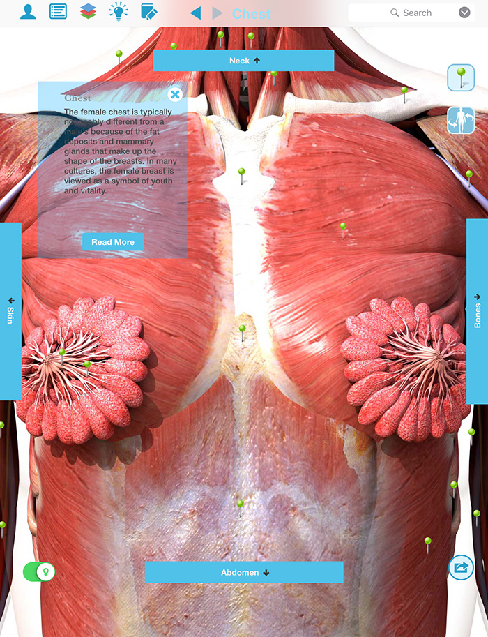
This page provides an overview of the chest muscle group.
Divides thoracic and abdominal cavities performs respiration. This muscle can be worked out in 2 different ways: The dominant muscle in the upper chest is the pectoralis major. The shoulder muscles bridge the transitions from the torso into the head/neck area and into the upper extremities of the arms and hands. It relieves tension in the chest and shoulders, and it also stimulates the abdominal organs, which helps to regulate digestion. Two sphincter muscles control the anus; This is the muscle that. Related online courses on physioplus. The transverse abdominal muscle wraps around the torso from front to back and from the ribs to the pelvis. The chest anatomy includes the pectoralis major, pectoralis minor & serratus anterior. The anterior muscles of the trunk (torso) are associated with the front of the body, include chest and abdominal muscles. Now that you have a basic understanding of what the abdominal muscles are and how they work, you can design workouts that actually target these muscles. Compress underlying structures rotate vertebral column rectus muscles:
The abdomen (colloquially called the belly, tummy, midriff or stomach) is the part of the body between the thorax (chest) and pelvis, in humans and in other vertebrates. Pigeon pose can feel intense and stimulating. In the hanging leg lift, the rectus abdominis must rotate the pelvis posteriorly and stabilize the pelvis to allow the legs to move freely toward the chest. This page provides an overview of the chest muscle group. Rectus abdominis, external abdominal oblique, internal abdominal it forms the bulk of the chest area and can be easily seen on the surface in some people, for the functions of the abdominal oblique muscles involve trunk flexion and ipsilateral rotation, as.
Chest Pictures Of Anatomy from www.sciencealert.com Flex vertebral column oppose erector spinae. Contraction of the diaphragm causes it to descend towards the abdomen, increasing the space of the thoracic cavity and expanding the lungs, filling them with air. This muscle forms the anterior and lateral abdominal wall. Compress underlying structures rotate vertebral column rectus muscles: Although the abdominal muscles have intersegmental nerve stimulation, you are not able to contract one section independent of the other. Hip flexion is the hip motion that brings the knee toward the chest. It relieves tension in the chest and shoulders, and it also stimulates the abdominal organs, which helps to regulate digestion. The anterior muscles of the trunk (torso) are associated with the front of the body, include chest and abdominal muscles.
The muscles of the abdomen also help with movement of the vertebral column and rotation of the trunk.
It enables the tilt of the pelvis and the curvature of the lower spine. Pigeon pose can feel intense and stimulating. Start studying chest and abdominal muscles. The internal sphincter, consisting of smooth muscle fibers, is under the control of the autonomic nervous system it is situated in the upper part of the abdominal cavity occupying the greater part of the right hypochondriac region, part of the epigastric region, and. This is the muscle that. The basic functions of these abdominal muscles involve providing structural support for the abdominal cavity as well as providing protection for the internal organs residing within the abdominal walls. Hip flexion is the hip motion that brings the knee toward the chest. Now that you have a basic understanding of what the abdominal muscles are and how they work, you can design workouts that actually target these muscles. Related online courses on physioplus. Divides thoracic and abdominal cavities performs respiration. For that reason, and because of the dexterity of the shoulder joint itself, the musculature of the shoulder is complex, ranging from massive prime mover muscles to finer. Muscles of the abdominal wall. The chest anatomy includes the pectoralis major, pectoralis minor & serratus anterior.
Fabian identifying the muscles and landmarks of the abdomen and chest chest muscles diagram. Hip flexion is the hip motion that brings the knee toward the chest.
Share :
Post a Comment
for "Chest And Abdominal Muscles Diagram ~ Anterior View of the Muscles of the Trunk | ClipArt ETC"


Post a Comment for "Chest And Abdominal Muscles Diagram ~ Anterior View of the Muscles of the Trunk | ClipArt ETC"